概念
・急性呼吸不全や低酸素血症の原因検索を迅速に行うための、ベッドサイドでの超音波検査法
・胸郭の上方と下方で、それぞれ前方、側方、後側方の6点から肺を観察する。
・肺炎に対して感度88%、特異度90%、心原生肺水腫に対して感度86%、特異度87%
観察部位
・胸郭の上方と下方で、それぞれ前方、側方、後側方の片肺計6点、両肺計12点から肺を観察する。
参照(このサイトより引用):https://ccforum.biomedcentral.com/articles/10.1186/s13054-021-03759-3
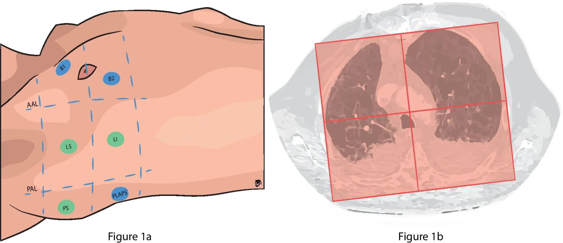
‘Bedside Lung Ultrasound in Emergency’ (BLUE) and 12-zone lung ultrasound protocol.
The blue points represent BLUE-points of the lung ultrasound protocol, they are identified as follows: two hands are placed consecutively just below the clavicle, excluding the thumbs. The upper BLUE-point is at the middle of the upper hand, whereas the Lower BLUE-point is at the middle of the lower palm. The posterolateral alveolar and/or pleural syndrome (PLAPS)-point is just below the posterior axillary line at the height of the diaphragm.
At the PLAPS-point the probe was moved posteriorly in order to obtain as much information as possible about the posterior lung region.
The green points are scanned additionally in the 12-zone lung ultrasound protocol. AAL: anterior axillary line; PAL: posterior axillary line; B1: BLUE-1; B2: BLUE-2; LS: lateral superior; LI: lateral inferior; PS: posterior superior; PLAPS: posterolateral alveolar and/or pleural syndrome.
b A schematic image of how thoracic CT was divided into two hemithoraces and an anterior and posterior region. Two lines were drawn tangent to the anterior and posterior border of both lungs and at the height of the sternum anteriorly and spinous process posteriorly a line perpendicular to the tangent lines was drawn to divide the thoracic cavity into a right and left side.
Both lungs were then divided into an anterior and posterior half by drawing a line perpendicular from the middle of the line between the sternum and spinous process to the lateral border of the lung. Each hemithorax was evaluated for anterior consolidations, posterior consolidations, interstitial syndrome, pneumothorax or pleural effusion
診るべき4つのポイント
参照:肺エコー(検査法と所見)
lung sliding
・臓側胸膜の呼吸性変動
・Bモードで観察
・気胸では消失(分かりにくい時は左右差を見るとよい)
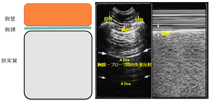
A line
・含気が良好な際に認められる水平方向の線状アーチファクト
・超音波画像のアーチファクトの代表的なものの一つである多重反射像が原因。
・多重反射像は、主としてプローブの振動子面と平滑な反射面との間を超音波が複数回往復することによって生じるアーチファクトです。
・肺エコーでは、プローブと平滑な胸膜との間を 1 往復して得られるのが真の胸膜の画像です。
・胸膜の下の肺実質には空気が多く含まれ、胸膜との音響インピーダンスの差による反射波が大きいことから、プローブの振動子面に戻った反射波の一部はもう一度生体内に押し戻されて胸膜での反射を 2 度、3 度と繰り返すことがあります。
・このようにプローブと胸膜との間を 2 往復、3 往復した超音波信号によって構成される多重反射像を A ラインと呼んでいます。
・プローブ面と胸膜との距離の 2 倍、3 倍の深さのところにあたかも胸膜の像のように表示される偽像が多重反射像(A ライン)の特徴です。
参照(このサイトより引用):https://www.fujifilm.com/jp/ja/healthcare/ultrasound/ultrasonography/ultrasound-column/general/lung-ultrasound
B line
・胸膜から画像深部まで減衰しないで伸びる彗星の尾のようなアーチファクト
・1肋間に3本以上見えるときに異常所見とする。
・肺水腫(前腋窩線第3肋間)、肺炎、ARDSなどで認める
PLAPS(posterolateral alveolar and/or plueural syndrome)
・肺が実質臓器のように観察される現象

コメント